On this page
What is glutathione?
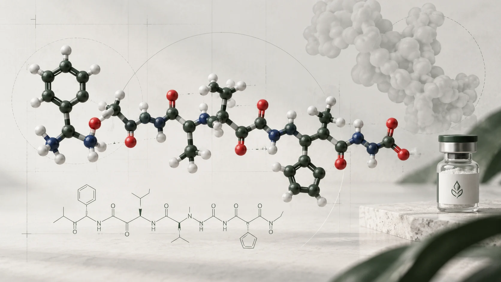
Glutathione is a tripeptide made from glutamate, cysteine and glycine. Its reduced form is usually abbreviated GSH, with the “SH” drawing attention to the free thiol group on cysteine. It occurs naturally in biological systems and is studied both as a chemical species and as part of cellular redox networks.
The name alone is not a complete description of a sample. Reduced glutathione, glutathione disulfide and glutathione attached to another molecule are different forms. A measurement described simply as “glutathione” may combine some of them or measure only one. Reading the method is therefore essential before comparing results.
Its scientific importance does not make every commercial claim about glutathione credible. A molecule’s role inside cells, a change in an experimental biomarker and a demonstrated human health outcome are separate questions. This Guide explains the chemistry and evidence, rather than proposing a treatment or attributing clinical findings to Lynx materials. For the broader terminology, see What Is a Peptide?
A tripeptide with an unusual linkage
Glutathione is written γ-L-glutamyl-L-cysteinyl-glycine. In most ordinary peptide backbones, the alpha-carboxyl group of one residue connects to the amino group of the next. Glutathione’s first connection instead uses the carboxyl group on glutamate’s side chain: the gamma-glutamyl linkage. The cysteine-to-glycine connection is an ordinary peptide bond.
This is a structural distinction, not an extra amino acid. There are still three residues. Writing Glu-Cys-Gly without indicating the gamma connection can leave out an important part of the identity. The account Glutathione homeostasis and functions describes that linkage and the molecular forms. The Peptide Sequence Guide explains why connections and modifications matter alongside residue letters.
Cysteine supplies a sulfur-containing thiol side chain. Changing its chemical state can alter glutathione without breaking either peptide bond. Conversely, cleavage of a peptide connection is a different chemical event. Structure therefore gives researchers a way to specify which change they are investigating, rather than treating every loss of starting material as the same reaction.
Reduced GSH and oxidised GSSG
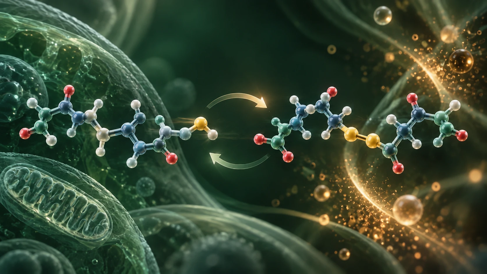
When two glutathione units are connected through their cysteine sulfur atoms, the product is glutathione disulfide, GSSG. It contains a disulfide bond between two peptide units. GSH and GSSG are related, but they are not interchangeable names for one unchanged compound.
Reduction of that disulfide can regenerate two GSH molecules. In cellular systems, glutathione reductase supports this conversion using reducing equivalents from NADPH. Glutathione peroxidase reactions can consume GSH while reducing suitable peroxides. These enzyme-coupled processes are more informative than the simple image of a peptide “absorbing toxins”.
Other forms also matter. Glutathione can form mixed disulfides with protein thiols or other small thiols, and it can participate in conjugation reactions. Glutathione in defense and signaling explains these different chemical routes. Measuring free GSH alone does not count every glutathione-containing species.
The definition of “total glutathione” should be stated. A method may reduce particular disulfides before detection, or combine measured pools mathematically. Whether a reported total counts GSSG molecules or glutathione equivalents affects the calculation. Two tables with the same heading can describe different measurement bases.
Redox chemistry is a system, not a single score
Oxidation and reduction involve electron transfer. In the GSH/GSSG couple, a change in sulfur chemistry connects those processes. Scientists use this relationship to investigate the conditions within a defined sample or cellular compartment. That is why glutathione appears so often in oxidative-stress research.
A GSH/GSSG ratio can be useful, but it does not describe the whole redox environment. Absolute concentrations, pH and the pool being sampled matter. Schafer and Buettner’s redox analysis explains why a ratio alone is insufficient to specify the couple’s reduction potential. A similar ratio at different concentrations need not describe the same chemical situation.
Cells also contain other redox systems. Their reactions are regulated and do not all sit at one shared equilibrium. The review of nonequilibrium thiol/disulfide systems discusses this context. A blood measurement cannot automatically describe a mitochondrial or nuclear pool, and a tissue extract can combine information from several compartments.
Oxidative stress is not simply “any oxidation”. Normal signalling includes controlled redox reactions. To argue that a system is under damaging stress, researchers need an appropriate experimental definition and supporting measurements, not just a changed glutathione value or an antioxidant label.
Synthesis, recycling and loss
Glutathione abundance reflects several processes at once. Cells synthesise it, recycle some oxidised forms, use it in reactions and transport or break down related species. A measured pool size is the net result, not a direct readout of any one of those processes.
Lu’s review of glutathione synthesis describes two enzyme-mediated steps: formation of gamma-glutamylcysteine, followed by addition of glycine. Precursor availability and enzyme regulation influence this pathway. The existence of cellular synthesis does not mean that adding a peptide to a culture will reproduce every aspect of endogenous control.
A study can investigate turnover by following labelled material, rather than measuring only concentration. Isotope-tracing research on glutathione and ophthalmate combined labelling with LC-MS/MS to investigate dynamics in its experimental systems. Pool size and synthesis or turnover rate answer different questions.
That distinction helps interpret apparently conflicting findings. An unchanged concentration can coexist with faster production and consumption. An increased concentration could reflect altered synthesis, reduced use, transport or a measurement artefact. A causal explanation needs a design that distinguishes those alternatives.
How laboratories study glutathione
Researchers may use chromatographic, mass-spectrometric, enzymatic or fluorescence-based methods. The choice depends on whether the question concerns free GSH, GSSG, related conjugates, total equivalents or change over time. Method selectivity is as important as sensitivity.
Paroni and colleagues used HPLC to distinguish glutathione fractions in biological samples. Nuhu and colleagues developed and validated an HPLC assay for GSH and GSSG. These are examples of defined analytical methods, not evidence that every assay or supplier report measures the same thing.
LC-MS can add mass-based selectivity, and appropriate internal standards can help account for variation in preparation and detection. Neither technology name guarantees complete recovery or correct assignment. The HPLC vs LC-MS Guide explains what separation and mass detection contribute.
Useful controls include blanks, reference materials, calibration and recovery assessment. Sample concentration, extraction and the reporting denominator also need attention. A result per volume, per cell count or per mass of tissue cannot be substituted for another without the relevant information. Comparing an experimental change requires the same measurement basis as well as a recognisable analyte name.
Oxidation during analysis can change the answer
Glutathione chemistry continues after sampling unless the method controls it. GSH can oxidise during preparation, making the measured reduced pool look smaller and the oxidised pool look larger. Such an artefact can resemble the biological change the experiment was designed to detect.
A primary investigation of blood GSSG measurements examined several sources of analytical distortion. Giustarini and colleagues’ methodological review shows why pre-analytical handling remains a central concern. These studies justify careful validation, not a universal handling recipe for all samples.
The timing and treatment of a sample should be documented alongside the result. Researchers also need to establish whether a method distinguishes the intended species after extraction. A tidy chromatogram does not prove that preparation preserved the original redox state.
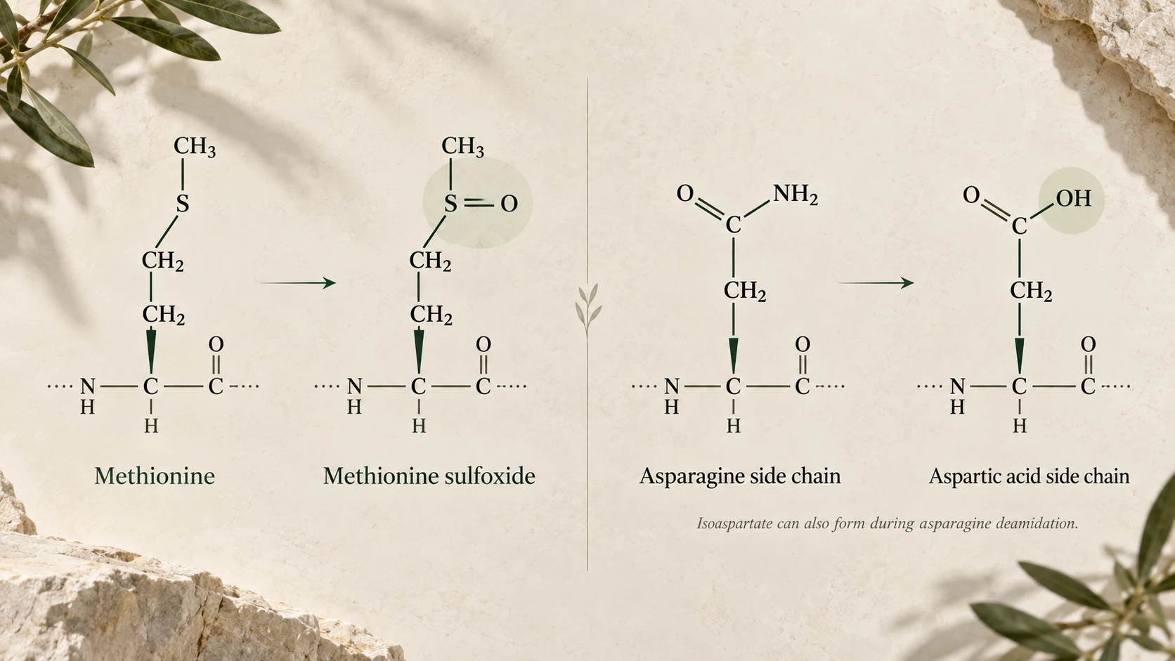
Physical recovery can create another problem. Material left on a container or excluded from the sampled fraction may change the measured amount without representing oxidation. The Solubility Guide distinguishes these physical questions. The Oxidation and Deamidation Guide explains why chemical changes require their own assignments.
What human research does and does not show
Human studies have investigated defined glutathione interventions, but they do not all ask the same question. A trial measuring a biomarker is not automatically a trial of symptoms, disease prevention or long-term health. The preparation, population, measurement and duration belong to the finding.
Allen and Bradley’s randomised placebo-controlled trial reported no significant changes in its measured oxidative-stress biomarkers or glutathione status in healthy adults. Richie and colleagues’ longer randomised trial reported increases in measured glutathione stores in specified sample types. Those results should be read with their respective designs, rather than reduced to “glutathione works” or “glutathione cannot work”.
A change in body stores does not itself establish a therapeutic outcome. Nor does a null biomarker result prove that every formulation and population will behave identically. Resolving a narrower clinical question requires trials designed to measure that outcome and assess harms.
Neither trial establishes an effect, safety or suitability of a Lynx research product. This article does not recommend supplementation or another route of use. Endogenous biochemical importance is a reason for investigation, not a licence to promise anti-ageing, detoxification or treatment benefits.
A practical framework for reading the evidence
Begin by identifying the chemical form and measurement. Then check what was changed experimentally and what was observed. This separates an association between glutathione and a condition from evidence that changing glutathione caused an outcome.
- Identify GSH, GSSG, mixed disulfides or a defined total.
- Check sample type, compartment and reporting units.
- Look for controls against oxidation during preparation.
- Distinguish concentration from turnover and reaction activity.
- Match conclusions to the model and measured endpoint.
- Keep biomarker findings separate from clinical outcomes.
For material documentation, the COA-reading Guide explains sample identity and test scope. Published biochemical research and a batch analytical report serve different purposes. One cannot fill gaps in the other.
Lynx materials are for laboratory research only, not for human or veterinary use, diagnosis or treatment. This article provides no dosing, administration or personal-use preparation instructions.
References and evidence scope
- Glutathione homeostasis and functions: potential targets for medical interventions. 2012. Structure and cellular context.
- Glutathione in defense and signaling: lessons from a small thiol. 2002. Enzyme and conjugation context.
- Schafer FQ and Buettner GR. Redox environment of the cell as viewed through the redox state of the glutathione disulfide/glutathione couple. 2001. DOI: 10.1016/S0891-5849(01)00480-4.
- Nonequilibrium thermodynamics of thiol/disulfide redox systems: a perspective on redox systems biology. 2008. Compartments and systems.
- Lu SC. Glutathione synthesis. 2013. DOI: 10.1016/j.bbagen.2012.09.008.
- Dynamics of glutathione and ophthalmate traced with 2H-enriched body water in rats and humans. 2009. Isotope-based turnover study.
- Paroni R et al. HPLC with o-phthalaldehyde precolumn derivatization to measure total, oxidized, and protein-bound glutathione in blood, plasma, and tissue. 1995.
- Nuhu F et al. Measurement of Glutathione as a Tool for Oxidative Stress Studies by High Performance Liquid Chromatography. 2020. DOI: 10.3390/molecules25184196.
- Blood glutathione disulfide: in vivo factor or in vitro artifact? 2002. Primary measurement study.
- Giustarini D et al. Pitfalls in the analysis of the physiological antioxidant glutathione (GSH) and its disulfide (GSSG) in biological samples: An elephant in the room. 2016. DOI: 10.1016/j.jchromb.2016.02.015.
- Allen J and Bradley RD. Effects of oral glutathione supplementation on systemic oxidative stress biomarkers in human volunteers. 2011. DOI: 10.1089/acm.2010.0716.
- Richie JP Jr et al. Randomized controlled trial of oral glutathione supplementation on body stores of glutathione. 2015. DOI: 10.1007/s00394-014-0706-z. Defined intervention, not Lynx evidence.