On this page
Chromatography and mass spectrometry answer different questions
Liquid chromatography separates components over time. Mass spectrometry measures ions according to their mass-to-charge ratio. Coupling the techniques in LC-MS connects a separation profile with mass information collected as material reaches the detector.
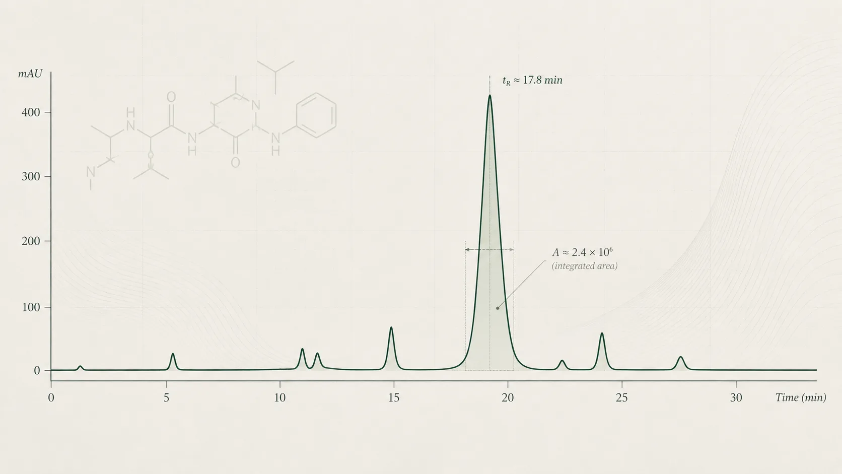
A chromatogram has a time axis. A mass spectrum has a mass-to-charge axis. A total ion chromatogram adds the recorded ion signal at each time point. An extracted ion chromatogram follows a selected mass-to-charge value or range. Neither should be confused with an HPLC-UV trace, which records a different detector response.
A clean chromatographic peak does not automatically contain one molecular species. Similarly, several peaks in a mass spectrum do not automatically mean several impurities. Read the separation and ion information together. The Waters mass-directed peptide isolation primer shows why complementary detectors can reveal different parts of a sample.
For the chromatographic measurement itself, see HPLC Peptide Purity Explained. This article concentrates on the identity information obtained from ions.
Molecular mass is not the same as mass-to-charge ratio
The neutral molecule and the ion entering a mass analyser are different entities. In positive electrospray, a peptide can acquire protons. The instrument then records the mass-to-charge ratio, written m/z, of the charged species.
For a protonated ion with charge number z, the relationship is m/z = (M + z × proton mass) ÷ z, where M is the neutral molecular mass. Rearranging gives M = z × m/z − z × proton mass. This expression applies to that specified ion, not to every possible adduct.
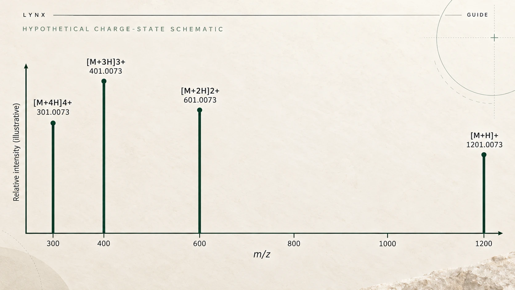
The notation [M+H]+ describes one added proton and a single positive charge. [M+2H]2+ describes two added protons and two positive charges. Comparing the second ion's m/z directly with the neutral mass would produce an incorrect conclusion.
Also check the mass convention. Monoisotopic mass uses specified isotope masses, while average mass reflects natural isotopic abundances. A calculated monoisotopic value and a reported average value are not interchangeable reference points. The report should make the convention and ion assignment clear.
Why one peptide can produce multiple charge states
A peptide can appear as several charged ions in the same electrospray spectrum. Charge distribution depends on molecular properties and analytical conditions. There is no universal rule that every peptide gives a strongest singly charged signal.
When ions assigned to one molecule occur at the same chromatographic retention time and give compatible reconstructed masses, they can support the same assignment. That agreement needs to be evaluated against the actual data. Co-elution alone does not prove that all recorded ions belong to one peptide.
Use the assigned charge states rather than simply counting spectral peaks. Missing or weak charge states can reflect detection conditions. They are not, on their own, evidence that a peptide is absent or structurally incorrect.
Isotope patterns, adducts and deconvolution
Natural isotopes create groups of related peaks around an ion signal. If the instrument resolves the isotope pattern, its spacing can help identify charge. A higher charge compresses the spacing on the m/z axis. This is different from a second charge-state group appearing elsewhere in the spectrum.
An adduct is another ion association, such as a sodium-containing species. Its mass relationship differs from the proton-only expression above. Assigning every signal as [M+H]+ can therefore create an apparent mass mismatch.
Deconvolution processes these charged patterns to estimate a molecular-mass representation. Thermo Fisher describes how its Xtract algorithm uses resolved isotope patterns and combines charge-state information. The output is processed evidence, not an additional independent experiment.
| Observation | Interpretation to check |
|---|---|
| A cluster of closely spaced peaks | An isotope pattern, with a charge assignment supported by resolution |
| Related clusters at different m/z values | Possible charge states of the same molecule |
| A displaced ion signal | A possible adduct, fragment or different component |
| A reconstructed mass peak | The result of the stated processing and assumptions |
Review the original spectrum alongside the processed result. Noise thresholds, mass ranges and overlapping signals can affect which components an algorithm reports. An uncluttered output is not proof that the original sample was equally simple.
Intact mass and fragmentation provide different evidence
An intact-mass measurement concerns the ionised molecule before deliberate fragmentation. Tandem mass spectrometry, often written MS/MS, selects a precursor ion and records product ions formed after fragmentation. The masses of those products can provide information about parts of the sequence.
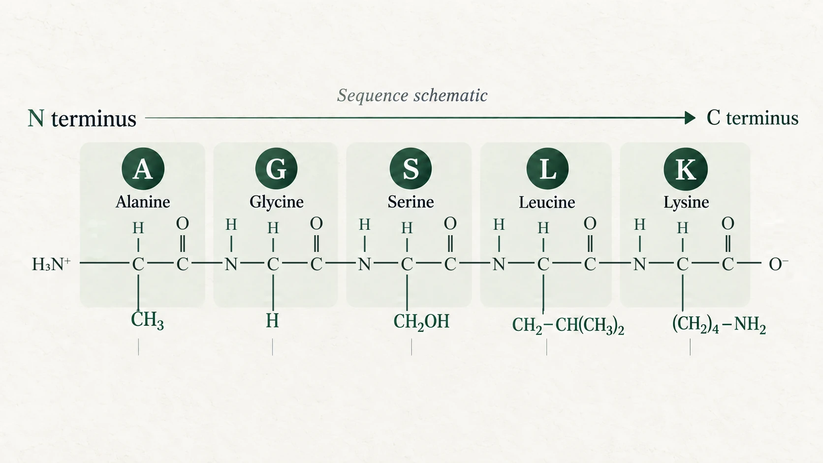
For common peptide fragmentation pathways, b-type ions retain the N-terminal side and y-type ions retain the C-terminal side. Other fragmentation methods produce different ion families. Thermo Fisher’s fragment-ion reference describes these conventions.
A useful sequence interpretation considers which expected fragments were observed, which positions remain unresolved and whether alternative assignments fit. Reporting that MS/MS was performed is not equivalent to demonstrating complete sequence coverage.
The precursor selection also matters. If more than one component contributes to the selected signal, the product-ion spectrum can contain contributions that complicate assignment. The method and supporting spectrum must justify the conclusion.
Why a matching mass does not establish complete sequence identity
Different residue orders can share the same overall composition and mass. Some residues are isomeric. Leucine and isoleucine, for example, have the same molecular mass, so an intact-mass match cannot distinguish them.
A 2014 study of Leu/Ile discrimination used multistage fragmentation to distinguish those residues in the investigated peptides. It demonstrates why a more specific question can require more specific evidence. It does not mean every routine LC-MS method provides that discrimination.
A molecular formula or mass value also does not establish stereochemistry. A report that supports a mass consistent with an expected peptide should not silently become a claim that every residue position, configuration and modification has been verified.
A report may express mass deviation in daltons or parts per million. Read the stated tolerance alongside the assigned ion and mass convention. A difference inside that tolerance supports the comparison made under the method, but it does not remove the structural ambiguities above. Do not infer an acceptance limit from the number of decimal places printed in a result. If the reference value, assignment or tolerance is missing, the strength of the reported comparison remains unclear.
Identity confidence depends on the question asked, method selectivity, comparison material and evidence supplied. Intact mass can be a useful part of that evidence without being a complete structural determination.
Reading an identity result responsibly
- Connect the sample. Establish which tested material the spectrum and result describe.
- Identify the measurement. Was the result an intact mass, a selected ion or fragmentation-based evidence?
- Read the assignment. Check charge, adduct and monoisotopic or average mass convention.
- Check comparison details. Look for the expected value, stated tolerance, calibration and reference approach.
- Review supporting data. Separate directly observed signals from software reconstructions and interpretations.
- Keep the conclusion bounded. Record unresolved sequence or stereochemical questions rather than assuming they were tested.
MS signal intensity alone is not a measured amount of peptide. Quantification needs an appropriate calibrated method. Equally, an identity result does not replace a chromatographic purity assessment. Understanding Peptide Analytical Testing explains how these separate measurement questions fit together.
Use the COA Guide for wider document checks and the QA library for published reports. The Research Library provides literature context, not identity evidence for a physical sample. This article describes analytical principles and does not claim that Lynx offers a particular MS method. Materials are for research use only, not for human or veterinary use.
References and further reading
- Waters: Considerations for Mass-Directed Purification. Laboratory primer on peptide ions, mass conventions and complementary detection.
- Discrimination of leucine and isoleucine in peptides sequencing with Orbitrap Fusion mass spectrometer. Primary research, 2014. PMID 24940639.
- Thermo Fisher: Understand the Xtract algorithm. Software reference for charge-state and isotope processing.
- Thermo Fisher: Fragment ions. Reference for peptide fragment-ion nomenclature.
Explore research materials
Browse research materials and consult the documentation for the relevant product and batch.